
Presentation
Patient was having a massage and felt shoulder pain - the concern was over whether there was a dislocation given the abnormal contour of the shoulder joint.
Patient Data

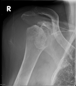
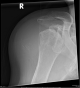
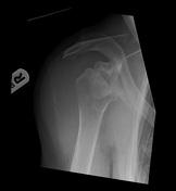
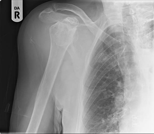
Severe arthropathy of the right shoulder with destructive changes to the humeral head and glenoid. Associated soft tissue swelling with calcification peripherally - these may represent bursal or intra-articular calcification. No convincing dislocation.
The appearances are highly suggestive of destructive arthropathy secondary to hydroxyapatite crystal deposition (Milwaukee shoulder).
Case Discussion
As seen in this case, Milwaukee shoulder frequently affects older women. Radiographic appearances largely resemble that of a neuropathic joint with destruction of the joint, soft tissue swelling and advanced rotator cuff tears. These patients are often managed conservatively.


 Unable to process the form. Check for errors and try again.