
Presentation
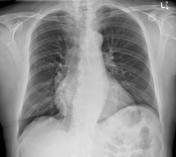
70 year old male presented with chest heaviness and shortness of breath. ECG showed atrial fibrillation.
Patient Data


Patient has longstanding mitral valve stenosis under review for mitral valve surgery.
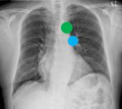
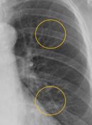
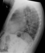
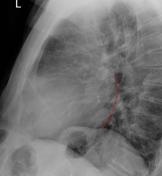
Left atrial enlargement is depicted by straightening of the left cardiac border (red) and a double right heart border (red). The remaining cardiac silhouette is outline in yellow (right atrium, right ventricle and left ventricle). Left atrial enlargement can also be appreciated on the lateral projection.
The cardiac silhouette is only mildly enlarged. Probable increase in pulmonary venous pressure can be inferred by comparing the size of the aorta knuckle with the left pulmonary artery at the aortopulmonary window. There equalization of the upper and lower lobe venous pressure.


 Unable to process the form. Check for errors and try again.