
Presentation
History of multiple falls with left leg trauma, most recent of which was 4 years ago, who presented with two days of left thigh pain
Patient Data


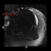
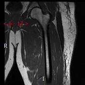
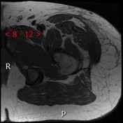
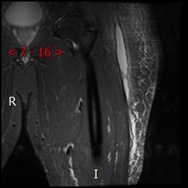
There is a 14.3 cm (CC) x 1.6 cm (transverse) x 5.2 cm (AP) low T1, high T2 collection between the subcutaneous fatty tissue and underlying fascia located over the greater trochanter of the left femur corresponding to the anechoic collection seen on the ultrasound and a density seen on the left hip radiograph. There is extensive overlying subcutaneous soft tissue edema. The findings are consistent with post-traumatic closed degloving/shearing injury (Morel-Lavallée lesion).
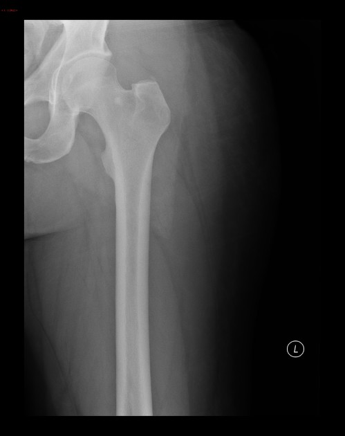
Large lobulated density projects over the subcutaneous soft tissues of the left thigh.
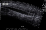
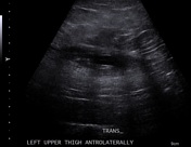
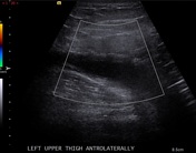
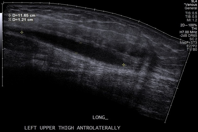
Mostly anechoic collection within the subcutaneous soft tissues of the left thigh without internal blood flow.
The collection was aspirated percutaneously. Gram stain and culture demonstrated rare WBCs, no growth.


 Unable to process the form. Check for errors and try again.