
Presentation
Painless, slowly increasing in size, post-traumatic right upper thigh mass for 5 months.
Patient Data
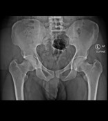
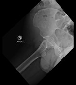
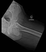

A well-defined homogeneous non-calcified soft tissue density is seen in the right upper lateral thigh. No underlying bone fracture or dislocation is seen.


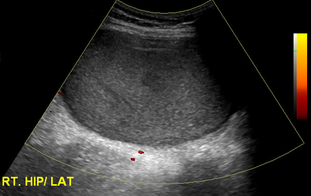
A well-defined heterogeneous fluid collection containing internal echoes/debris, is seen in the subcutaneous soft tissues of the right upper lateral thigh. No internal vascularity is seen in it.
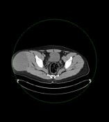

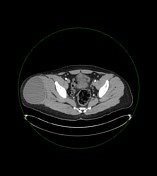
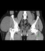
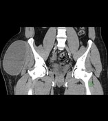
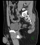
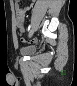
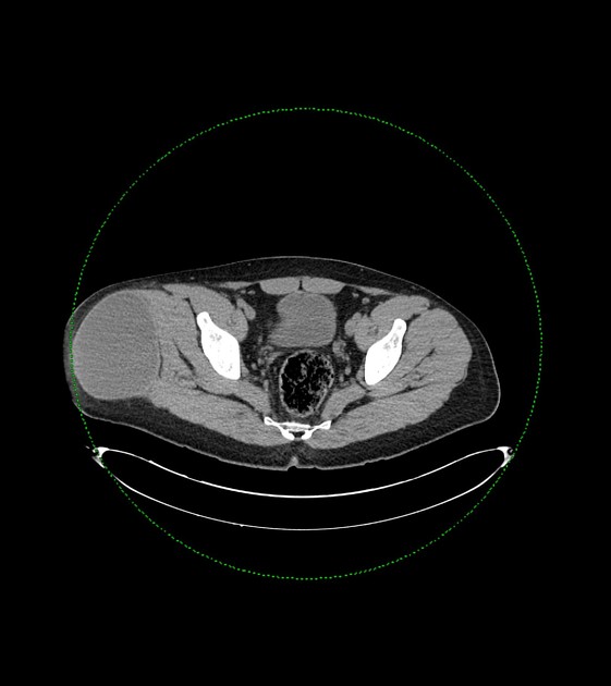
A well-defined encapsulated cystic lesion measuring 9 x 10 x 15 cm (AP x Trans x CC), having an average density of 33 HU, showing minimal peripheral enhancement, is seen in the subcutaneous soft tissues of the right upper lateral thigh, superficial to the tensor fascia lata. No septations, fat/solid component or internal enhancement is seen in it.
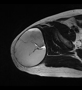
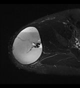
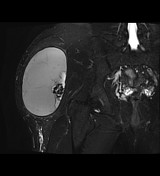
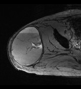
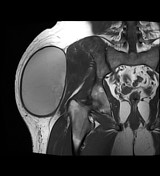
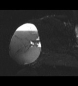
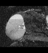
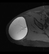
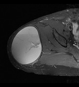
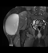

A well-defined encapsulated cystic lesion is seen in the subcutaneous soft tissues of the right upper lateral thigh, superficial to the tensor fascia lata. It is hyperintense on both T1 & T2-weighted images, and has fluid-fluid level and multiple thin septations. No fat/solid component or internal enhancement is seen in it.
Well-defined cystic lesion in the subcutaneous soft tissues of the right upper lateral thigh, superficial to the tensor fascia lata. In view of history of trauma, this lesion is likely a Morel-Lavallée lesion.
Case Discussion
The patient underwent right gluteal hematoma evacuation and debridement.
Gross description: The specimen is received in formalin in one container, labeled with the patient's name, medical record number and "right gluteal sac". It consists of a previously opened cystic lesion measuring 15 x 10 x 9 cm and weighing 311 grams. The cystic wall thickness is 0.3 cm. One surface is smooth and glistening and the other surface shows focal hemorrhagic areas with fatty tissue.
Microscopic description: Pseudocyst showing zones of fibrin, dense fibrosis, granulation tissue, foci of lymphohistiocytic inflammation & hemorrhage consistent with Morel-Lavallée lesion.
Case courtesy of Dr Ahmed Eid


 Unable to process the form. Check for errors and try again.