
Presentation
History of trauma 2 weeks ago, now right thigh swelling.
Patient Data


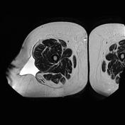
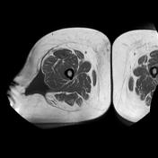
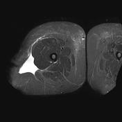
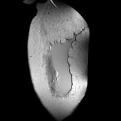
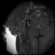
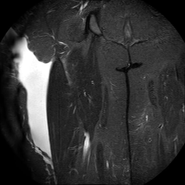
Large well defined fluid collection within the deep subcutaneous plane of the lateral aspect of the right thigh.It's overlying the tensor fascia lata and vastus lateralis muscle.
It appears hypointense on T1 WI and hyperintense on T2 WI with internal fat lobules are seen entrapped within the lesion.
The lesion measures about 8.0 x 5.0 x 22 cm along its max TS, AP and CC dimensions.
Case Discussion
A Morel-Lavallée lesion represents a closed degloving injury associated with severe trauma which then presents as a hemolymphatic mass.
MRI is the imaging modality of choice for evaluation.
Early diagnosis and management is essential to avoid complications such as infection or extensive skin necrosis.


 Unable to process the form. Check for errors and try again.