
Presentation
History of trauma.
Patient Data
Age: 40 years
Gender: Female
From the case:
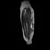
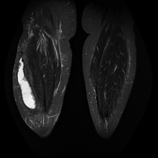
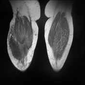
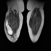
Morel-Lavallée lesion - leg

Download
Info

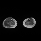
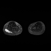
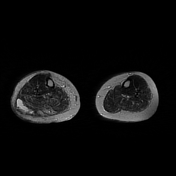
Large well defined fluid collection within the deep subcutaneous plane of the lateral aspect of the right leg.
Diffuse subcutaneous soft tissue edema of the right leg.
Case Discussion
Morel-Lavallée lesions are closed degloving injuries associated with severe trauma which then present as hemolymphatic masses.
MRI is useful for the diagnosis.
The lesions typically occur over the greater trochanter of the femur, yet, similar biomechanical forces may occur at the lumbar region, over the scapula, the knee, or the leg 1 can result in identical Morel-Lavallée lesions.


 Unable to process the form. Check for errors and try again.