
Presentation
Right knee trauma. Persistent pain.
Patient Data
Age: 35 years
Gender: Female


Download
Info
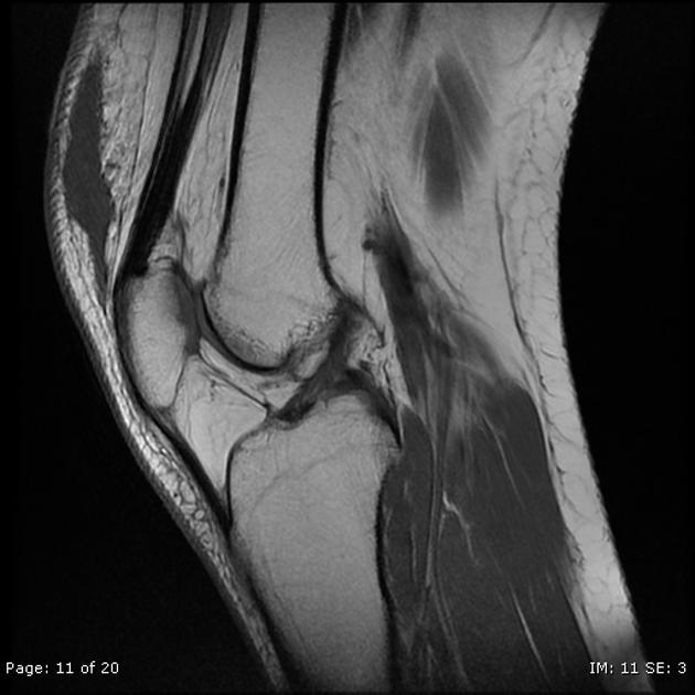
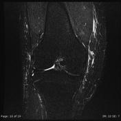
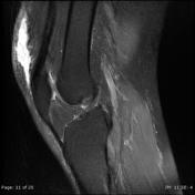
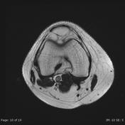
A 7 x 7 x 1.7 cm subcutaneous elliptical fluid collection eliciting low T1 and high T2/PDFS/STIR signals is seen anterosuperior to the patella enclosed within the splitted deep fascia.
Altered marrow signal of posterior aspect of medial tibial condyle eliciting low T1 and high T2/PDFS/STIR signal.
Minimal intra-articular joint effusion.
Case Discussion
Morel-Lavallée lesion (closed degloving injury) with marrow edema/contusion of the posterior medial tibial condyle.
Although Morel-Lavallée lesion is a name given to this lesion when it occurs over the greater trochanter, similar lesions can occur over the knee due to a similar mechanism.


 Unable to process the form. Check for errors and try again.