

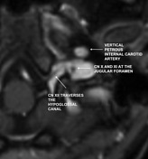
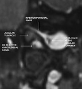
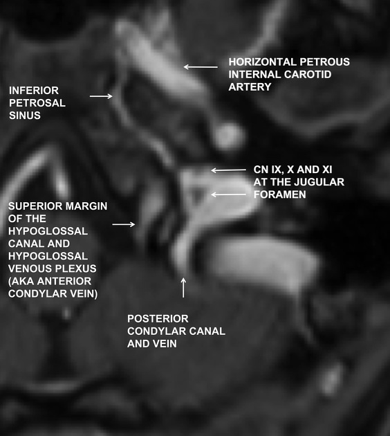
Gadolinium enhanced MPRAGE in the axial (Fig A and B) and coronal (Fig C) planes illustrate the anatomy of cranial nerves IX, X, XI and XII. These cranial nerves are surrounded by rich venous networks at the skull base and contrast enhanced MRI sequences provide the best contrast resolution to image the pathway of these cranial nerves through the skull base.
Cranial nerve IX, X and XI traverse through the pars nervosa and pars vascularis portion of the jugular foramen which is positioned superolateral to the jugular tubercle. In contrast, cranial nerve XII lies within the hypoglossal canal inferomedial to the jugular tubercle. On the coronal plane the jugular tubercle assume the morphology of the "eagle's head". Erosion or remodeling of the jugular tubercle, "eagle's head", is a characteristic feature of neoplastic pathology arising from the jugular foramen.


 Unable to process the form. Check for errors and try again.