Presentation
Epigastric pain and swelling 1 year. No vomiting, weight loss or change in bowel habits.
Patient Data
![](https://prod-images-static.radiopaedia.org/images/54884336/8_thumb.jpeg)
![](https://prod-images-static.radiopaedia.org/images/54884337/9_thumb.jpeg)
![](https://prod-images-static.radiopaedia.org/images/54884338/10_thumb.jpeg)
![](https://prod-images-static.radiopaedia.org/images/54884335/7_thumb.jpeg)
![](https://prod-images-static.radiopaedia.org/images/54884333/2_thumb.jpeg)
![](https://prod-images-static.radiopaedia.org/images/54884336/8_big_gallery.jpeg)
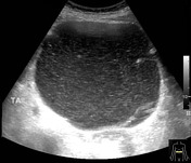
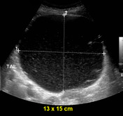
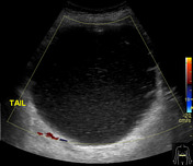
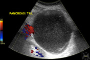
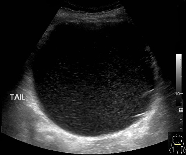
A large well-defined cyst containing turbid contents and multiple thin septations, measuring 13 x 15 cm, likely arising from the tail of the pancreas, is seen in the epigastric region. No solid component or significant internal vascularity is appreciable in it.
![](https://prod-images-static.radiopaedia.org/images/54884544/Anonymize.Seq2.Ser3.Img95_thumb.jpeg)
![This study is a stack](/packs/stack-YQKLCKBI.gif)
![](https://prod-images-static.radiopaedia.org/images/54884619/Anonymize.Seq3.Ser7.Img95_thumb.jpeg)
![This study is a stack](/packs/stack-YQKLCKBI.gif)
![](https://prod-images-static.radiopaedia.org/images/54884488/Anonymize.Seq4.Ser200.Img31_thumb.jpeg)
![This study is a stack](/packs/stack-YQKLCKBI.gif)
![](https://prod-images-static.radiopaedia.org/images/54884644/Anonymize.Seq6.Ser600.Img31_thumb.jpeg)
![This study is a stack](/packs/stack-YQKLCKBI.gif)
![](https://prod-images-static.radiopaedia.org/images/54884804/Anonymize.Seq5.Ser201.Img39_thumb.jpeg)
![This study is a stack](/packs/stack-YQKLCKBI.gif)
![](https://prod-images-static.radiopaedia.org/images/54884892/Anonymize.Seq7.Ser601.Img38_thumb.jpeg)
![This study is a stack](/packs/stack-YQKLCKBI.gif)
![](https://prod-images-static.radiopaedia.org/images/54884544/Anonymize.Seq2.Ser3.Img95_big_gallery.jpeg)
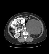
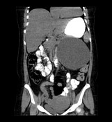
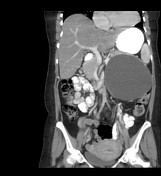
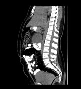
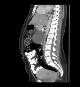
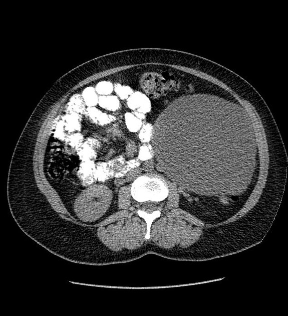
Homogeneous well-defined cystic lesion measuring 14.5 x 14.5 cm, arising from the pancreatic tail displacing the adjacent structures. Thin enhancing wall, multiple thin septae and a few tiny peripheral calcifications are seen in it. No solid component is seen. Bulky postpartum uterus.
Impression: Large homogeneous well-defined pancreatic tail cystic lesion, which is likely a mucinous cystadenoma; other possible differential can be a pancreatic pseudocyst if there is any history of pancreatitis.
Case Discussion
Procedure: Distal pancreatectomy and splenectomy.
Gross Description: The pancreatic cystic lesion measures 16 x 14 x 13 cm and weighs 161 grams.
Diagnosis: Mucinous cystadenoma of the pancreas, measuring 16 cm.