
Presentation
Memory problems.
Patient Data
Note: This case has been tagged as "legacy" as it no longer meets image preparation and/or other case publication guidelines.
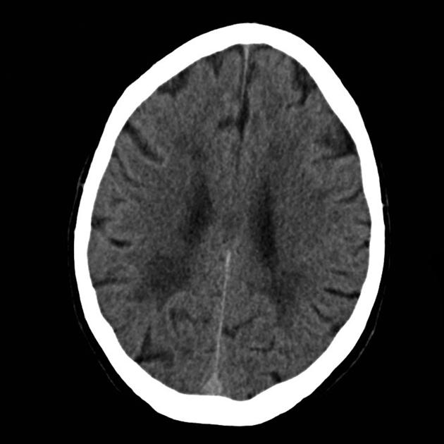
Single axial non-contrast CT image demonstrates patchy periventricular hypodensity seen bilaterally.

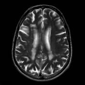
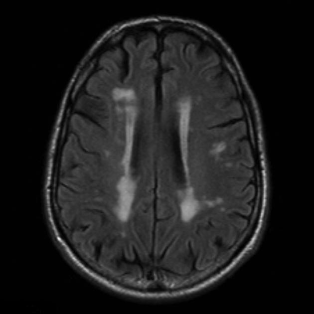
MRI demonstrates multiple T2 white matter hyperintensities seen on both T2 and FLAIR images.
Case Discussion
These features are non-specific and common and represent chronic small vessel ischemic change. It used to be thought that Alzheimer's disease and vascular dementia were separate, if not exactly mutually exclusive. We now recognize that they often co-exist. As such before a diagnosis of vascular dementia can be made, a careful assessment for imaging and neurocognitive features of Alzheimer's disease should be undertaken.


 Unable to process the form. Check for errors and try again.