
Presentation
The patient originally underwent an MRI for headaches. A pineal cyst was detected, that was followed up for several years.
Patient Data
Age: 30 years
Gender: Female
Note: This case has been tagged as "legacy" as it no longer meets image preparation and/or other case publication guidelines.
From the case:
Multiloculated pineal cyst
Download
Info
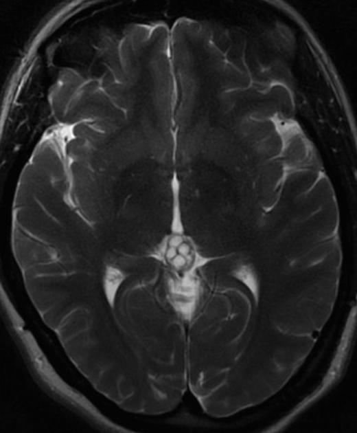
T2-weighted MRI showed a thin-walled, multiloculated cyst in the pineal region, that did not demonstrate interval growth over a period of three years.
Case Discussion
Pineal cysts are common and when typical in appearance they require no follow-up. Larger cysts or those with atypical features (such as this one) may need to be followed up to ensure they do not represent cystic pineal neoplasms (e.g. pineocytoma).


 Unable to process the form. Check for errors and try again.