
Presentation
Altered sensorium.
Patient Data
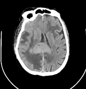

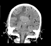
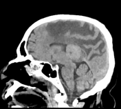

Multiple well-defined homogenously hyperdense lesions involving the right frontal lobe, left inferior frontal lobe, and splenium of the corpus callosum with marked surrounding edema.

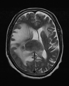
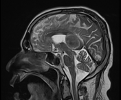
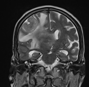
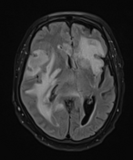
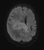
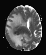
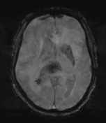
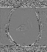

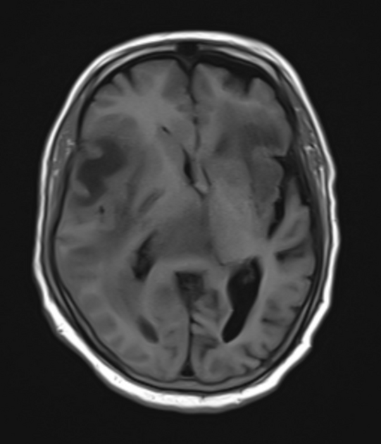
Multiple well-defined rounded mass lesions involving the right frontal lobe, left inferior frontal lobe, and splenium of the corpus callosum with marked surrounding edema. These lesions appear iso to hypointense on T1 weighted images, and hypointense on T2/FLAIR. High signal on DWI and very low ADC values, consistent with pronounced diffusion restriction. Microhemorrhage are seen on SWI. Vivid homogeneous enhancement on T1 post-contrast images.
Case Discussion
Overall imaging findings are highly suggestive of multiple CNS lymphoma. Unfortunately the patient passed away prior to biopsy.
Co-author: Dr. Avijit Kashyap (Consultant Neurosurgeon).


 Unable to process the form. Check for errors and try again.