
Presentation
Manifested in early teens with epileptic seizures, general weakness, progressive cognitive decline, emotional lability, shaky writing, and walking. Tonic-clonic seizures were successfully treated with a combination of two antiepileptic drugs, but the eradication of myoclonic status wasn't achieved. The patient also has hypothyroidism and cardiomyopathy.
Patient Data


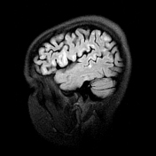
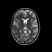
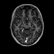
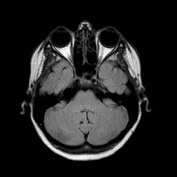
MRI scans have shown predominantly cortical hyperintense lesions on T2 and FLAIR sequences in both cerebral hemispheres, basal ganglia, midbrain, and cerebellum. T1 hypointensities in the corresponding areas with no contrast enhancement.
MRI scans and laboratory testing (aciduria, decrease of plasma carnitine, increase of blood and CSF lactate, etc.) suggested that the patient might have mitochondrial pathology. Genetic analysis has shown the mutation in the MT-ND5 gene. The latter is characteristic of MERRF syndrome, MELAS, and Leigh syndrome, of which MERRF syndrome was the most likely diagnosis, judging by the clinical picture. Unfortunately, the patient refused to perform a muscle biopsy to confirm the diagnosis.
Case Discussion
This case shows both the radiological and clinical picture of MERRF syndrome . Primary mitochondrial disorders are especially hard to diagnose properly, so a thorough differential diagnosis is required, relying on both on MRI and genetic analysis.


 Unable to process the form. Check for errors and try again.