
Presentation
Incidental finding during work-up for dysphagia and gastro-esophageal reflux disease.
Patient Data
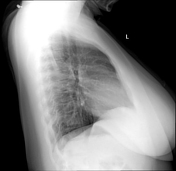
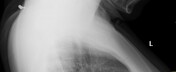
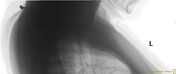

An example of a nape piercing with a subcutaneous bar and two surface balls.
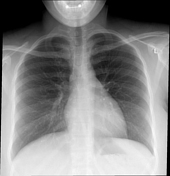
The lung fields are clear. There is a normal cardiothoracic ratio. There are no indirect features to suggest a hiatal hernia.
Case Discussion
An example of a nape surface piercing. The initial frontal view may falsely suggest a possible oral cavity, lip, or tongue piercing. The lateral view confirms otherwise and demonstrates a nape piercing with a subcutaneous metal bar and two visible surface balls.
Surface piercings usually have an entrance and exit hole while standard piercings usually have a solitary hole.
The patient was unable to remove the piercing for the X-rays. This was physically attempted in our department after the piercing was identified.


 Unable to process the form. Check for errors and try again.