
Presentation
Decreased hearing on the right.
Patient Data
Note: This case has been tagged as "legacy" as it no longer meets image preparation and/or other case publication guidelines.

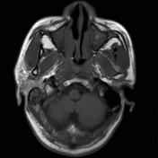
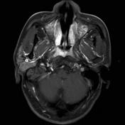
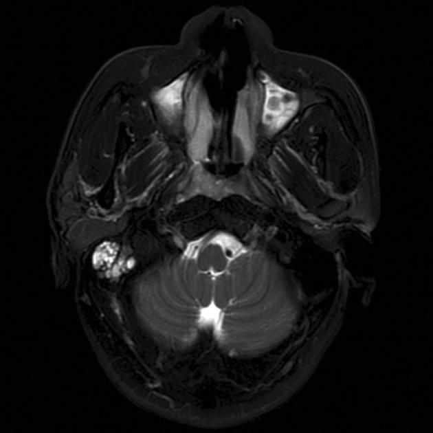
Selected images demonstrates a poorly defined mass involving the right side of the nasopharynx, with increased T2 and enhancement. It is associated with fluid in the mastoid air cells.
The patient went on to have a biopsy.
Histology
The section shows lymphoid tissue covered on one surface by respiratory type epithelium. Within the lymphoid tissue there are irregularly shaped islands and trabeculae of a densely hypercellular tumor.
Tumor cells have large irregularly shaped vesicular nuclei and a moderate amount of pale eosinophilic cytoplasm and are arranged in diffuse sheets. Moderate numbers of mitotic figures are noted. Intercellular bridges are discernible in some areas of the tumor. No evidence of keratinization is seen.
Tumor cells show strong immunostaining for cytokeraqtins (AE1/AE3; CAM5.2) and epithelial membrane antigen (EMA). There is also patchy weak staining for chromogranin. No staining for EBV-LMP is seen. The features of poorly differentiated non-keratinizing squamous cell carcinoma.
Final diagnosis: Poorly differentiated squamous cell carcinoma.


 Unable to process the form. Check for errors and try again.