
Presentation
Term neonate admitted to hospital with urinary tract infection. Ultrasound was requested to rule out underlying pathology.
Patient Data
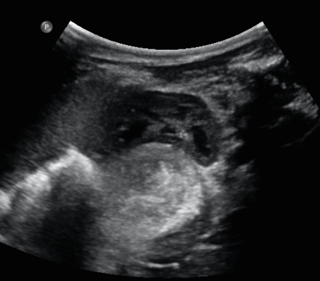
Spot images from renal ultrasound representing a mass posterior to left kidney hilum.
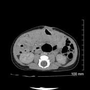

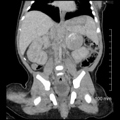
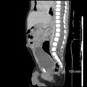
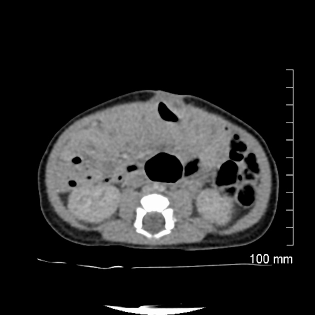
Large, partially peripherally calcified mass is seen anteriorly to the upper pole of the left kidney corresponding to the findings from ultrasound and representing neuroblastoma (confirmed histopathologicaly).
Case Discussion
Neonatal neuroblastoma is a type of congenital neuroblastoma, which is diagnosed within the first month of the life. In the majority of cases, the tumour is localised in the adrenal gland.
Neonatal neuroblastoma accounts for less than 5% of all cases of neuroblastoma and carries a favourable prognosis with most cases being low risk for metastases and recurrence, however around 20% of neonatal neuroblastoma presents with spinal cord compression due to metastatic disease.
This case presents neuroblastoma arising from the left adrenal gland which could have been easily mistaken for stomach or loop of bowel on ultrasound.


 Unable to process the form. Check for errors and try again.