
Presentation
The patient presented with epilepsy, she had an ex-brother known to have ceroid neuronal lipofuscinosis, and she was recently diagnosed.
Patient Data
Age: 4 years
Gender: Female
From the case:
Neuronal ceroid lipofuscinosis


Download
Info
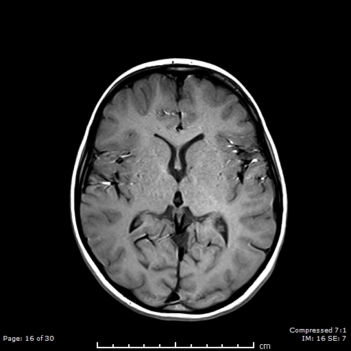
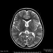
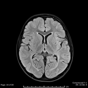
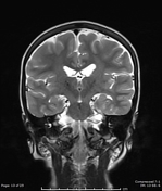
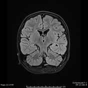
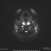
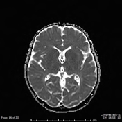
Bilateral periventricular symmetric T2/FLAIR hyperintensity is noted, along with mild periventricular leukoencephalopathy and mild cerebral atrophy.
The thalami exhibit T2 hypointensity.
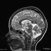
Prominent folia of the cerebellar hemispheres, representing cerebellar atrophy.
Case Discussion
Imaging findings of neuronal ceroid lipofuscinosis are non-specific; however, the presence of cerebellar and/or cerebral atrophy, leukoencephalopathy and thalamic T2W-hypointensity along with clinical presentation are highly suggestive of the diagnosis of NCL 1.
T2 hypointensity can also be seen in gangliosidosis.


 Unable to process the form. Check for errors and try again.