
Presentation
Two weeks of foot swelling and redness, not responsive to antibiotics.
Patient Data


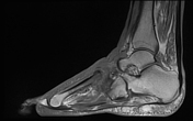
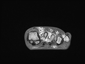
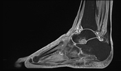
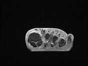
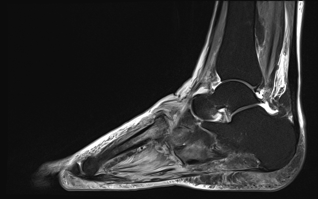
Abnormality centred on the tarsometatarsal joint, where there is extensive subluxation without dislocation and arthropathic changes with disrupted joint spaces, subchondral oedema and enhancement with only mild low T1 bone marrow signal.
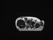
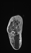
Extensive cutaneous and subcutaneous thickening, oedema and enhancement.
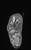
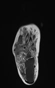
Intrinsic muscles of the foot demonstrate diffuse oedema and enhancement with relatively sparing of the lumbricals and interossei in the forefoot. No regions of non-enhancement to suggest necrosis. Less vivid oedema of the muscles in the visualised lower leg muscles.
Case Discussion
Findings are in keeping with a neuropathic (Charcot) joint noting chronically elevated HbA1c (10+ years in the poorly controlled or very poorly controlled range) results. In this setting, the muscle changes are in keeping with diabetic myopathy.


 Unable to process the form. Check for errors and try again.