
Presentation
Headache, expressive dysphasia, altered consciousness. Fevers, chills, rigors. Recent travel from Uganda.
Patient Data


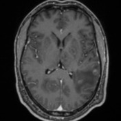
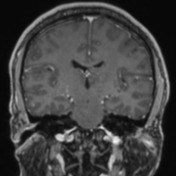
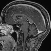
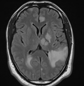
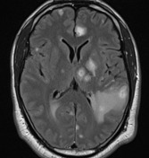
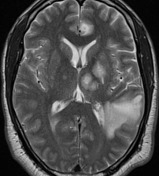
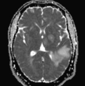
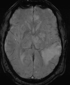
Innumerable, irregular, peripherally contrast enhancing lesions are seen in the supra and infratentorial brain, of varying sizes. These are mostly distributed in the basal ganglia and at the grey-white matter junction. The largest are located in the parietal lobes, measuring up to 4.5 cm on the right and 2.7 cm on the left. Several of lesions have a mural nodule e.g. in the right basal ganglia, and the left frontal lobe lesion has a target-like pattern of enhancement. No convincing central diffusion restriction. Many are however T2 hypointense centrally, with foci of susceptibility. Extensive surrounding vasogenic edema. No definite leptomeningeal or pachymeningeal thickening/hyperenhancement.
No ventricular enlargement or ependymal enhancement to suggest ventriculitis. No subfalcine, uncal or tonsillar herniation. Paranasal sinuses are unremarkable.
Case Discussion
The patient subsequently underwent a craniotomy and biopsy, which confirmed the presence of toxoplasmosis gondii on microscopy and immunohistochemistry. Patient was also newly diagnosed with HIV, with HIV-related immunosuppression thought to be the predisposing factor for neurotoxplasmosis. MRI features suggestive of neurotoxoplasmosis include:
- peripherally-enhancing lesions of varying sizes distributed in the basal ganglia and grey-white matter junctions
- extensive vasogenic edema surrounding the lesions
- several lesions have a mural nodule e.g. right basal ganglia
- the left frontal lobe lesion has a target-like pattern of enhancement, which can be seen in neurotoxoplasmosis


 Unable to process the form. Check for errors and try again.