Presentation
Right sided neck swelling.
Patient Data
Age: 20 years
Gender: Female
From the case:
Nodular fasciitis of the neck
![](https://prod-images-static.radiopaedia.org/images/62439207/971e2902bd1ecf5e0c34ac47c9d0b94e2c420dd36b9cb3faa415f0b29161fe25_thumb.jpeg)
![This study is a stack](/packs/stack-YQKLCKBI.gif)
![](https://prod-images-static.radiopaedia.org/images/62439307/099541b6257f35466b463f709e54452de62716395cc7680f028761ddd65dd97f_thumb.jpeg)
![This study is a stack](/packs/stack-YQKLCKBI.gif)
![](https://prod-images-static.radiopaedia.org/images/62439219/186ada3944edaae9fd716c039bd183f72bf7a7c613190e866506b31aac956314_thumb.jpeg)
![This study is a stack](/packs/stack-YQKLCKBI.gif)
![](https://prod-images-static.radiopaedia.org/images/62439250/e1eccb0ccd558e157874998501c2f4c30e584b4ee5888163b05d7d60e439dd74_thumb.jpeg)
![This study is a stack](/packs/stack-YQKLCKBI.gif)
![](https://prod-images-static.radiopaedia.org/images/62439269/5b92dc756ed84e49f978888ddc3162463cff425630bd4f71bfa5ed76cca59293_thumb.jpeg)
![This study is a stack](/packs/stack-YQKLCKBI.gif)
![](https://prod-images-static.radiopaedia.org/images/62439289/f14f42ee85736b2f92a76eab6aead6aba876a47e9ca2646ef17e62b004c2038a_thumb.jpeg)
![This study is a stack](/packs/stack-YQKLCKBI.gif)
Download
Info
![](https://prod-images-static.radiopaedia.org/images/62439207/971e2902bd1ecf5e0c34ac47c9d0b94e2c420dd36b9cb3faa415f0b29161fe25_big_gallery.jpeg)
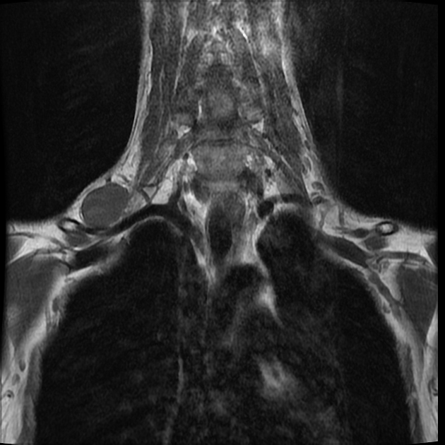
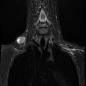
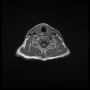
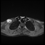
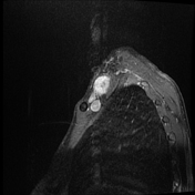
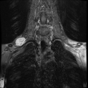
Right supra-clavicular well defined mass lesion related to right brachial plexus shows isointense signal in T1 and heterogeneous hyperintense signal in T2 with avid enhancement on post-contrast study.
Case Discussion
Histopathology and immunohistochemistry showed features of nodular fascitis.
Possible differential in this case include nerve sheath tumor (schwannoma, neurofibroma) due to close proximity to brachial plexus.