
Presentation
Painful distal left thigh
Patient Data
Age: 9 years
Gender: Female
From the case:
Non-ossifying fibroma
Download
Info
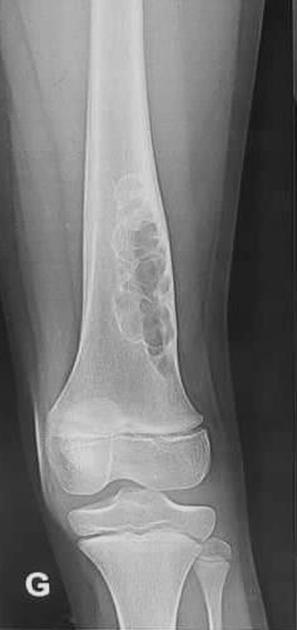
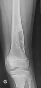
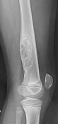
There is an expansile eccentric multiloculated lucent lesion with peripheral sclerotic rim, involving the posterolateral aspect of the diaphysometaphyseal region of the distal femur. No pathological fracture or periosteal reaction is seen.
From the case:
Non-ossifying fibroma


Download
Info
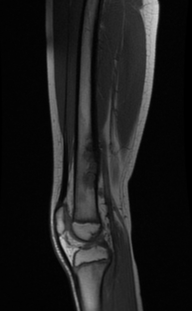
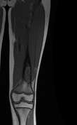
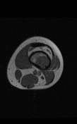
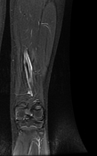
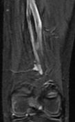
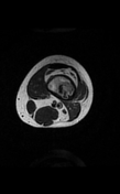
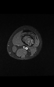
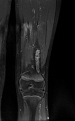
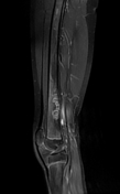
The MRI sequences demonstrate a multiloculated cortical-based lesion of intermediate signal on T1WI, heterogeneous high signal on T2WI with peripheral low signal rim corresponding to the sclerotic border. The postcontrast sequences show moderate heterogeneous enhancement. No soft tissue mass or edema is seen.
Case Discussion
Radiographic and MRI features are most consistent with a non-ossifying fibroma of the distal femur.
Additional contributor: C. Boukaaba, MD


 Unable to process the form. Check for errors and try again.