Presentation
Young patient with psychotic presentation - organic cause?
Patient Data
![](https://prod-images-static.radiopaedia.org/images/17420180/78de707f66bbf12bf2da188b96bb2e_thumb.jpeg)
![This study is a stack](/packs/stack-YQKLCKBI.gif)
![](https://prod-images-static.radiopaedia.org/images/17420069/d68403f51ebc76d0fee3f220425b88_thumb.jpeg)
![This study is a stack](/packs/stack-YQKLCKBI.gif)
![](https://prod-images-static.radiopaedia.org/images/17419225/cbdd1322bbd7805d92cffb279eacd2_thumb.jpeg)
![This study is a stack](/packs/stack-YQKLCKBI.gif)
![](https://prod-images-static.radiopaedia.org/images/17419563/6eba6214de4eb2ece328258e9a3946_thumb.jpeg)
![This study is a stack](/packs/stack-YQKLCKBI.gif)
![](https://prod-images-static.radiopaedia.org/images/17419812/7c5976508221679b044caae4a0e8c3_thumb.jpeg)
![This study is a stack](/packs/stack-YQKLCKBI.gif)
![](https://prod-images-static.radiopaedia.org/images/17420456/3fe39ee5938e66cd5e7cdf9f01237a_thumb.jpeg)
![This study is a stack](/packs/stack-YQKLCKBI.gif)
![](https://prod-images-static.radiopaedia.org/images/17420326/b8a54424ea4e41fb2f871b21001f1c_thumb.jpeg)
![This study is a stack](/packs/stack-YQKLCKBI.gif)
![](https://prod-images-static.radiopaedia.org/images/17420180/78de707f66bbf12bf2da188b96bb2e_big_gallery.jpeg)
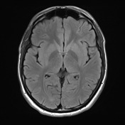
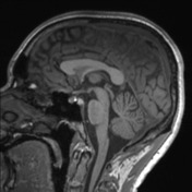
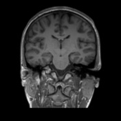
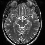
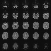
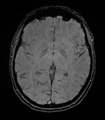
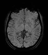
Technique: Multiplanar and multisequence imaging has been obtained through the brain including T1 and isotropic acquisition.
Findings: Appearance and intensity of brain parenchyma are normal. Ventricular system and cisternal spaces appear normal. No evidence of intracranial space occupying lesion or obvious vascular anomaly is detected. The visualized orbits, paranasal sinuses and calvarium appear unremarkable.
Conclusion: Normal exam.
Case Discussion
This case illustrates a normal Brain MRI in an 18-year-old boy. Please, refer to the article on normal brain imaging examples for more cases like this. Note that absence of the occipital horns is a normal variant.