
Presentation
Chronic left wrist pain
Patient Data
Age: 35 years
Gender: Male


Download
Info

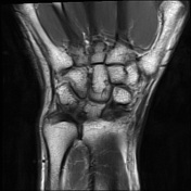
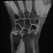
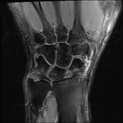
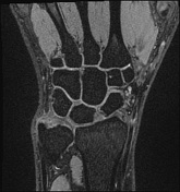
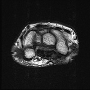
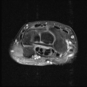
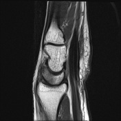
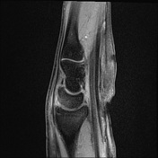
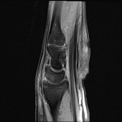
Neutral ulnar variance. The triangular fibrocartilage including the disc proper is intact. Ulnomeniscal homologue is intact. Extensor carpi ulnaris tendon is intact.
Scapholunate ligament is intact. Bone shows normal signal intensity. No joint effusion.
Case Discussion
This is a normal left wrist MRI. The injury suspected is at TFCC (triangular fibrocartilage complex), however, the TFCC is normal. This is an example of MRI 3 Tesla of the wrist without an arthrogram.
"T2 de3D we" is a sequence specifically for Siemens MRI machine. It is also known as DESS (Double Echo Steady State). It is commonly used in musculoskeletal imaging and can be MPR into 3D imaging for assessment.


 Unable to process the form. Check for errors and try again.