
Presentation
Headache.
Patient Data
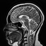

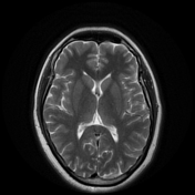
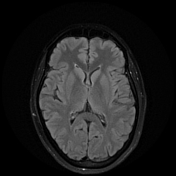
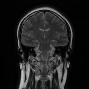
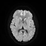
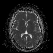


Normal MR signal of the cerebral hemispheres, cerebellum, and brainstem. Normal size and signal of the ventricular systems. No atrophic or hydrocephalic changes. No extra-axial collections. No midline shift.
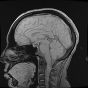
Morphologic examination of the aqueduct of Sylvius showed normal dimensions of the duct with no evidence of obstruction or pathological dilatation.
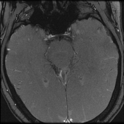
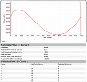
Qualitative assessment of the cine-phase examination showed free CSF flow signal through the aqueduct of Sylvius, foramina of the fourth ventricle, and around the brain stem of the foramen magnum. Quantitative assessment showed:
- systolic mean flow = 0.007 mL/s
- duration of CSF systole = 450 ms
- stroke volume = 3.5 microliter (normal up to 42 microliter)
Case Discussion
The MR CSF flowmetry results are in keeping with normal CSF flow dynamics with normal CSF quantitative assessment and morphological features showing no evidence of obstruction.


 Unable to process the form. Check for errors and try again.