
Presentation
Reduced sensation of L4/L5/S1 regions
Patient Data
Age: 20 years
Gender: Female


Download
Info

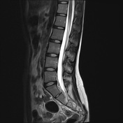
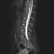
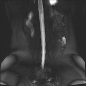
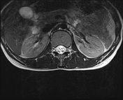
Last unfused vertebra is taken as L5. Normal lumbar lordosis and spinal alignment. Vertebral body heights are preserved. The intervertebral discs shows normal signal intensity. Spinal cord ends at the level of T12, with normal signal of the visualized spinal cord.
No disc bulge. Spinal canal are spacious at all these levels. The lateral recesses and exit foramina are spacious at all these levels. No traversing or exiting nerve root impingement. No ligamentum flavum hypertrophy. No facet joint arthropathy.
Case Discussion
This is a normal MRI of the lumbosacral spine.


 Unable to process the form. Check for errors and try again.