
Normal posterior meniscofemoral ligament - ligament of Wrisberg
Presentation
Knee pain
Patient Data


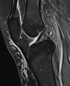
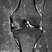

MRI study demonstrates prominent posterior meniscofemoral ligament (ligament of Wrisberg).
Medial femoral condyle osteochondral lesion.
Degeneration of the posterior horn of the medial meniscus.
Mild knee joint effusion.
Baker's cyst showing small loose bodies.
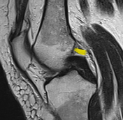
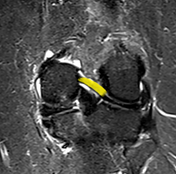

Annotated images follow the course of the posterior meniscofemoral ligament (ligament of Wrisberg).
Case Discussion
The meniscofemoral ligament (MFL) arises from the posterior horn of the lateral meniscus and passes to attach to the lateral aspect of the medial femoral condyle. It splits into two bands at the posterior cruciate ligament (PCL), which are named in relation to the PCL:
- anterior meniscofemoral ligament (ligament of Humphrey)
- posterior meniscofemoral ligament (ligament of Wrisberg)
Radiologists should be aware of the normal course of the MFL, not to be misinterpreted on sagittal MR images as an intra-articular loose body or meniscal fragment.
A special meniscal tear is known as Wrisberg rip also occurs at the junction of the ligament of Wrisberg and the posterior horn of the lateral meniscus and is commonly associated with anterior cruciate ligament tears.


 Unable to process the form. Check for errors and try again.