
Presentation
Cognitive decline, incontinence and gait disturbance.
Patient Data


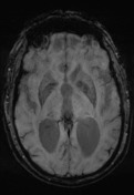
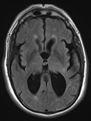

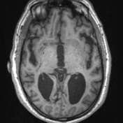
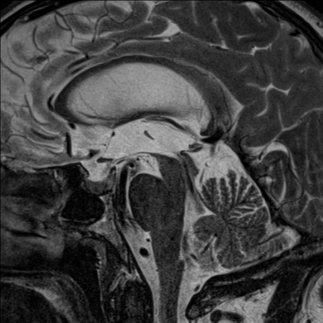
Multiple FLAIR hyperintensities scattered throughout the periventricular and subcortical white matter likely reflect chronic small vessel ischaemia, prominent in severity for this age. Even allowing for a degree of exvacuo dilatation related to this, the lateral and third ventricles are prominent in size compared to the sulcal pattern and the corpus callosum is thinned.
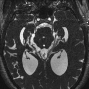
There is disproportionate crowding of the sulci superiorly near the vertex and widening of the sylvian fissures. The flow void is exaggerated across the aqueduct of Sylvius. Patency is also demonstrated on the CSF flow study (not shown)
Conclusion:
Appearances compatible with normal pressure hydrocephalus.
Case Discussion
A diagnosis of NPH on imaging alone is difficult and care must be taken to correlate with patient symptoms. Much of the initial work done on CSF flow studies has been difficult to replicate.


 Unable to process the form. Check for errors and try again.