Normal shoulder arthrogram

Case contributed by Matt Skalski
Diagnosis not applicable

Patient Data

Age: 21
Gender: Female
mri
This study is a stack
Axial T1
C+ fat sat
This study is a stack
Coronal T1
C+ fat sat
This study is a stack
Sagittal T1
C+ fat sat
This study is a stack
Coronal
T1 C+
This study is a stack
Coronal T2
fat sat
Download
Info

Unremarkable MR arthrogram of the shoulder. 

MR arthrogram coronal oblique

Annotated image
posterior section -
infraspinatus muscle
middle section -
glenoid cavity
anterior section -
biccipital tendon
Download
Info
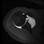
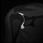
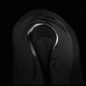
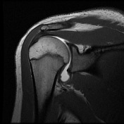
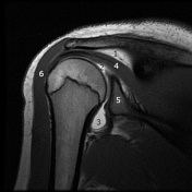
  1. subdeltoid-subacromial fat

  2. contrast outlining the articular surface of the humerus

  3. inferior glenohumeral ligament forming the axillary pouch

  4. infraspinatus muscle

  5. glenoid fossa

  6. deltoid muscle

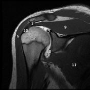
  7. superior glenoid labrum

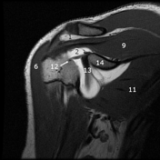
  8. inferior glenoid labrum

  9. supraspinatus muscle

  10. insertion supraspinatus tendon into the greater tuberosity of the humerus

  11. subscapularis muscle

  12. tendon of the long head of biceps brachii

  13. anterior glenoid rim

  14. coracoid process

Case Discussion

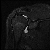
This is a good example of a normal MR arthrogram of the shoulder. 

How to use cases

You can use Radiopaedia cases in a variety of ways to help you learn and teach.

Creating your own cases is easy.

:

Updating… Please wait.

 Unable to process the form. Check for errors and try again.

 Thank you for updating your details.