
Presentation
Long standing left facial pain, worse with eating meals
Patient Data
Age: 35
Gender: Male
From the case:
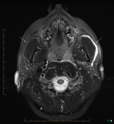
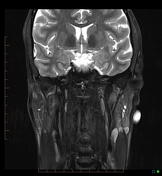
Obstructed parotid duct: MR sialogram

Download
Info
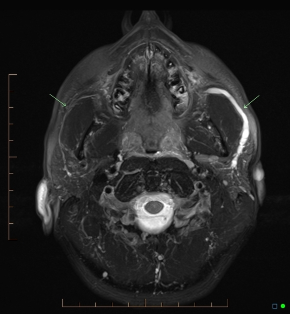
Dilated left parotid duct from the gland extending up to the left cheek where there is a tapered narrowing. Note the asymmetry when compared with the right (arrows). Note also the dilated accessory duct entering the main duct (best seen on the sagittal imaging)
From the case:
Obstructed parotid duct: MR sialogram

Download
Info
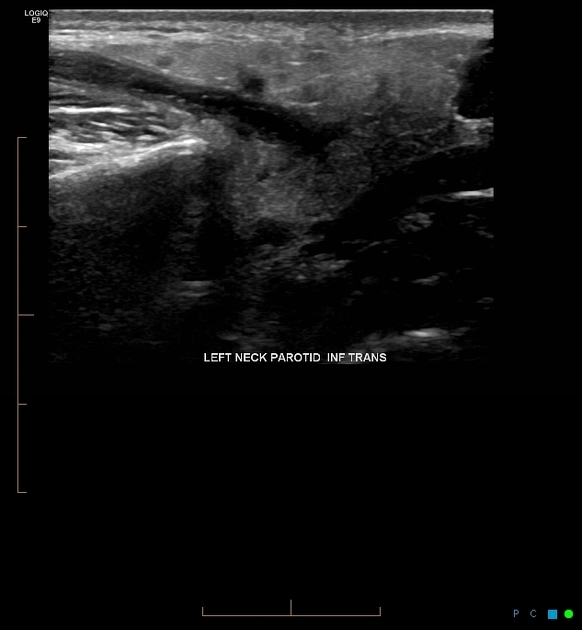
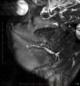
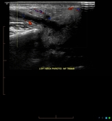
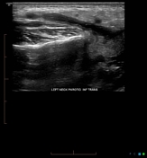
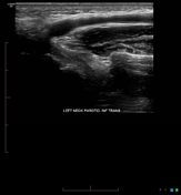
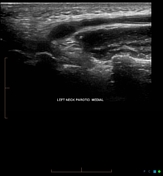
US confirms the MRI findings and shows minor increase in vascularity in the gland parenchyma. No intra-luminal calculus at the distal end of the duct in the cheek.
Case Discussion
Catheter sialography is fiddly and uncomfortable for the patient. MR sialography is a useful alternative especially if the ducts are dilated


 Unable to process the form. Check for errors and try again.