Presentation
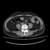
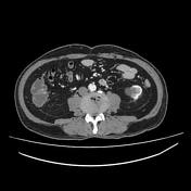
Recurrent left renal colic.
Patient Data
Age: 75 years
Gender: Male
From the case:
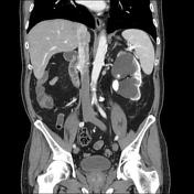
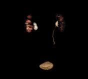
Obstructive uropathy





Download
Info
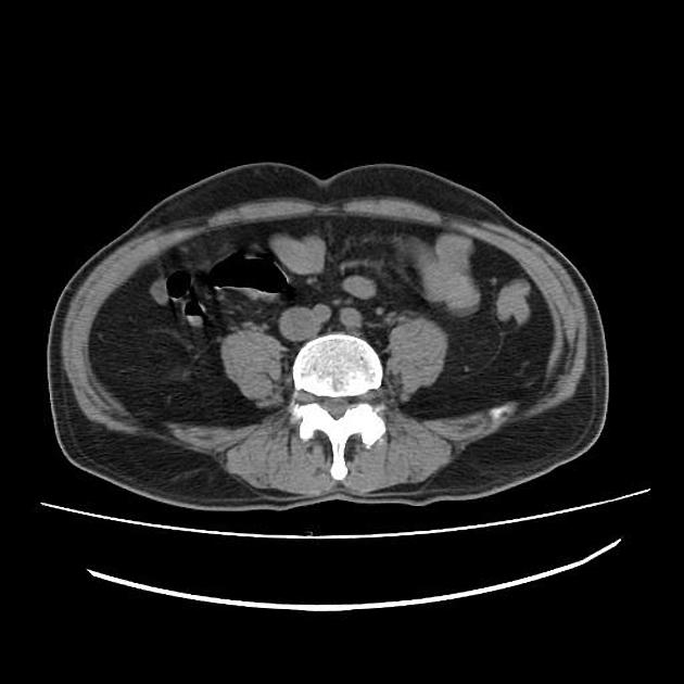
The left kidney shows:
- marked hydronephrosis with thinning of the cortical renal parenchyma and delayed contrast excretion on the excretory phase
- calyceal stones ranging from 6 to 10 mm (mean density = 720 HU)
- retracted renal pelvis with two contiguous obstructive stones measuring 17 and 25 mm (mean density = 1400 HU)
- two simple cortical renal cysts measuring 15 and 65 mm
- normal caliber of the ureter with no ureteric stone is seen
Right kidney shows
- normal size and shape
- normal renal cortical thickness
- normal appearance of the calyceal system and renal pelvis
- no renal stone
- small simple cortical renal cyst is noted (17 mm)
- normal caliber of the ureter
The urinary bladder shows a normal appearance with no bladder stone is seen
Other findings:
- biliary microlithiasis (<3 mm in diametre)
- prostatic hypertrophy (volume = 45 mlsc)
- aortic atherosclerosis
Case Discussion
CTU features of obstructive uropathy due to renal pelvis stones with reduced renal cortical thickness and delayed excretion on excretory phase.


 Unable to process the form. Check for errors and try again.