
From the case:
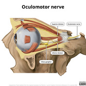
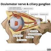
Oculomotor nerve and ciliary ganglion (illustration)

Download
Info

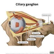
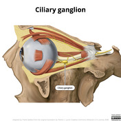
Illustration of the orbit with the lateral wall removed demonstrating the course and branches of the oculomotor nerve as well as the ciliary ganglion.
Case Discussion
These illustrations are adapted from the original illustration by Patrick Lynch released on Creative Commons Attribution 2.5 License 2006 and available on Wikimedia commons here.
A number of changes were made:
- optic nerve made lighter to distinguish from oculomotor nerve
- supply to superior oblique muscle removed (this is supplied by the trochlear nerve, not the oculomotor nerve)
- ciliary ganglion simplified
- superior oblique passes under superior rectus (modified by Anka Friedrich here)


 Unable to process the form. Check for errors and try again.