Presentation
Seizure.
Patient Data


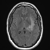
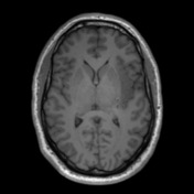
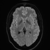
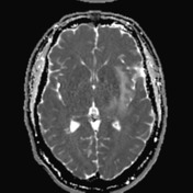
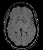
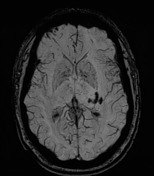
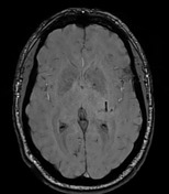
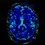
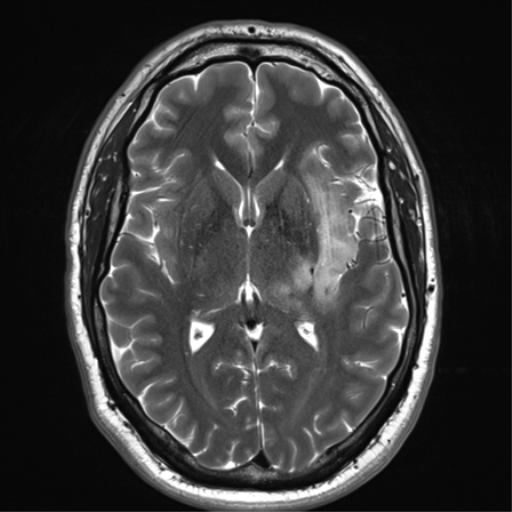
Left-sided ill-defined high T2 signal left insular lesion involving cortex and subjacent white matter extending up into the periventricular white matter of the posterior left frontal lobe is again demonstrated. Embedded within the lesion are multiple areas of coarse calcification. Some intrinsic high T1 signal is present, along with some vessels, but no convincing parenchymal enhancement. MR spectroscopy (not shown) demonstrates elevation of choline and mild loss of. Cerebral blood volume is not elevated. ADC demonstrates facilitated diffusion.
Conclusion: Appearances are characteristic of a diffuse glioma, and the presence of calcification and morphological features strongly suggest that this represents an oligodendroglioma.
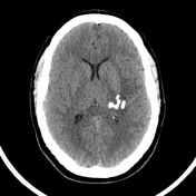
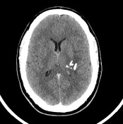

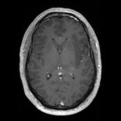
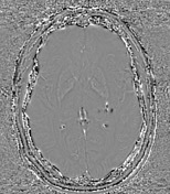
Calcific densities in the left hemisphere surrounded by hypodensity with positive mass effect.
Case Discussion
The patient went on to have a resection.
Histology
Sections show mildly to moderately hypercellular fragments of brain tissue infiltrated by tumor cells with optically clear cytoplasm and centrally located round to ovoid nuclei with evenly distributed chromatin. Infiltrative growth including neuronal satellitosis is seen. There is no microvascular proliferation or palisading necrosis. Mitoses are rare.
Immunohistochemistry:
- GFAP Positive
- IDH-1 R132H Positive (mutated)
- ATRX Positive (not mutated)
- p53 Mosaic pattern (probably not mutated)
- p16 CDKN2A Mosaic pattern (probably not mutated)
- Ki67 labeling index: Approximately 5%
Mollecular markers:
- 1p/19q codeletion: DETECTED
- EGFR amplification: Not detected
- IDH1 codon 132: PATHOGENIC VARIANT DETECTED (R132H)
- IDH2 codon 172: No pathogenic variant detected
- BRAF codon 600: No pathogenic variant detected
- H3F3A codons 27 and 34: No pathogenic variant detected
- TERT promoter Chr5:1295228 (C228T) and chr5:1295250 (C250T): PATHOGENIC VARIANT DETECTED (C228T)
FINAL DIAGNOSIS: Oligodendroglioma, IDH-mutated and 1p/19q-codeleted (WHO grade II).


 Unable to process the form. Check for errors and try again.