
Presentation
Patient is referred for an obstetric ultrasound at term.
Patient Data

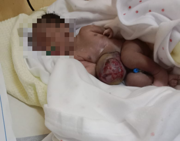
Post-partum photo of the baby a few hours later after vaginal delivery shows the anterior abdominal wall defect as seen on ultrasound.
Consent obtained to share image.
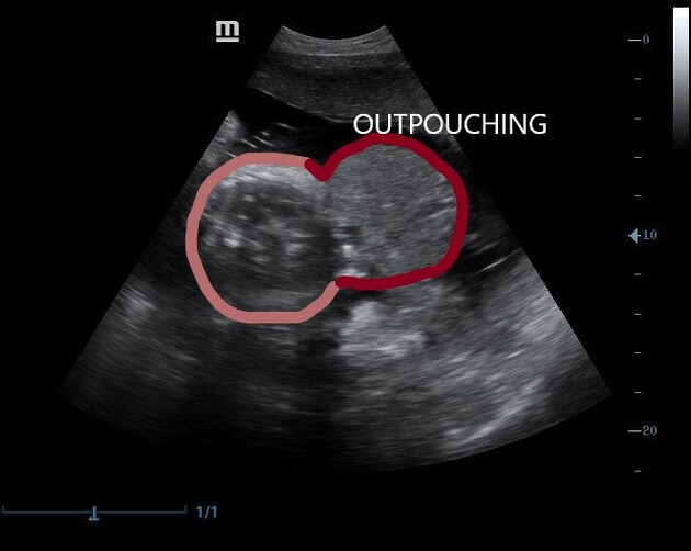
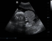
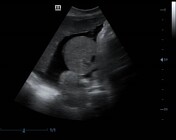
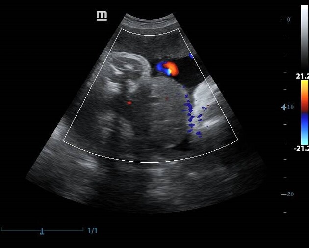
Illustrated image showing normal abdominal wall outline and the membrane-covered defect.
Case Discussion
Omphalocele is a congenital mid-line wall defect through which abdominal viscera herniate into a membrane-covered sac. It has been suggested that the formation of an omphalocele may be due to failure of the two lateral embryonic wall folds to fuse during embryologic development at 3-4 weeks. Due to the higher likelihood of related anomalies, smaller omphaloceles are believed to have a worse prognosis.


 Unable to process the form. Check for errors and try again.