
Presentation
Left orbital pain with partial visual loss.
Patient Data
Age: 40 years
Gender: Male
From the case:
Optic neuritis


Download
Info
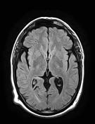
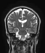
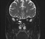
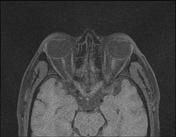
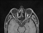
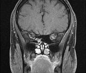
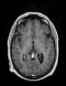
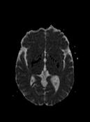
The MRI sequences demonstrate a left unilateral swelling of the retrobulbar intra-orbital segment of the optic nerve extending to the intracranial segment, of high T2 signal with enhancement on postcontrast sequences.
Normal appearance of the right optic nerve and optic chiasma.
Case Discussion
MRI features of left optic neuritis.


 Unable to process the form. Check for errors and try again.