Presentation
Known patient with lung cancer presented with bilateral proptosis.
Patient Data
![](https://prod-images-static.radiopaedia.org/images/63115434/83856e225096187dd84721de76fceba7e21ecf24a716d12cb2c9d53624294747_thumb.jpeg)
![This study is a stack](/packs/stack-YQKLCKBI.gif)
![](https://prod-images-static.radiopaedia.org/images/63115457/83d40297fc337489417f5effa547565d88d2a426981b866d52741e805c7f6ea9_thumb.jpeg)
![This study is a stack](/packs/stack-YQKLCKBI.gif)
![](https://prod-images-static.radiopaedia.org/images/63115243/fc3a754b200f2bdaaec74c676da59acb2d8a01b45950752506b3001f95cf996b_thumb.jpeg)
![This study is a stack](/packs/stack-YQKLCKBI.gif)
![](https://prod-images-static.radiopaedia.org/images/63115205/637d43343284885bd0784aaf908cac67468fa547058b5916fe6aafde3867e6db_thumb.jpeg)
![This study is a stack](/packs/stack-YQKLCKBI.gif)
![](https://prod-images-static.radiopaedia.org/images/63115308/6a2edee1bf163209684893fd287bc427b109b81ff9fbab4bae3d3a4bca394220_thumb.jpeg)
![This study is a stack](/packs/stack-YQKLCKBI.gif)
![](https://prod-images-static.radiopaedia.org/images/63115254/a8980f7d8a9b6935d1849c64a0abefe86ac92cd9c9da6a4bec16e604765612b8_thumb.jpeg)
![This study is a stack](/packs/stack-YQKLCKBI.gif)
![](https://prod-images-static.radiopaedia.org/images/63115354/9a7da3fe8bece75d5c5ed72ba771a6626fcc5dd8b869decd9e25716f02b27a0e_thumb.jpeg)
![This study is a stack](/packs/stack-YQKLCKBI.gif)
![](https://prod-images-static.radiopaedia.org/images/63115331/d596aa746053583ca639700c24a3194ab89a8d17272faa48b19ba3f35b6342d0_thumb.jpeg)
![This study is a stack](/packs/stack-YQKLCKBI.gif)
![](https://prod-images-static.radiopaedia.org/images/63115396/75a46ae778c40010bf6e44fbe3c1caa1e4cbc1eda06af4eebe2499051aa5abf2_thumb.jpeg)
![This study is a stack](/packs/stack-YQKLCKBI.gif)
![](https://prod-images-static.radiopaedia.org/images/63115434/83856e225096187dd84721de76fceba7e21ecf24a716d12cb2c9d53624294747_big_gallery.jpeg)
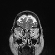
Bilateral proptosis.
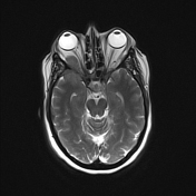
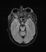
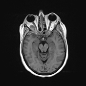
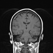
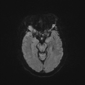
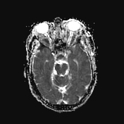
Both orbits show extraconal masses with isointense T1 and T2 signals, larger on right orbit, encroaching upon extraocular muscles. They show diffusion restriction and post-contrast enhancement. On right side, they are extending to orbital apex compressing the optic nerve in optic canal.
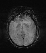
Diffuse pachymeningeal thickening and enhancement, with more thickening at right parietal, left squamous temporal and bilateral anterior temporal regions, more on right side.
Altered texture and signal intensity of diploic spaces at both posterior parietal regions ? skull lesions.
Case Discussion
The patient has a known history of lung cancer. Bilateral orbital lesions and the presence of dural thickening are impressive of metastatic disease.