
Orbital apex mass - primary squamous cell carcinoma of sphenoid sinus
Presentation
Deteriorating vision right eye.
Patient Data


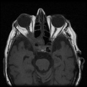
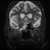
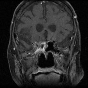
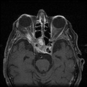
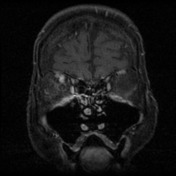
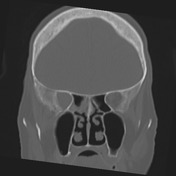
MRI study shows a soft tissue enhancing mass involving the cavernous sinus, the right sphenoid and soft tissue extending into the orbit. It surrounds the optic nerve and does not arise directly from it.
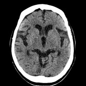
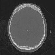
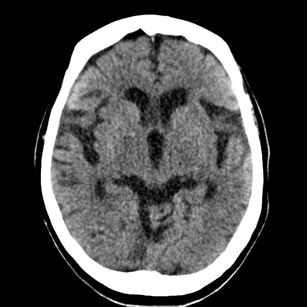
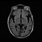
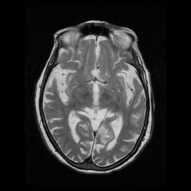
There is significant involutional change in the brain. There is a mass lesion arising in relationship to the anterior portion of the right cavernous sinus, this is associated with destructive change in the lateral wall of the adjacent right sphenoid air cell.
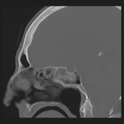
There is expansion of the superior oblique fissure of the right orbit and the tumour gains access to the right orbital apex as a consequence. There is also some bony destruction in the region of the base of the right anterior clinoid close to the right optic canal.
There is evidence of otomastoiditis on the right and one wonders if there is some erosion in the region of the Aditus. There is, nonetheless, no significant right nasopharyngeal soft tissue abnormality and the fossa of Rosenmuller is clear on both sides.
CONCLUSION:
Mass lesion in the region of the right cavernous sinus extending towards and possibly into the right sphenoid air cell and extending through the superior oblique fissure into the apex of the right orbit. The mass is indistinguishable from the margins of the right optic canal as well.
Case Discussion
The soft tissue mass involving the right cavernous sinus, the sphenoid air cells, and the orbital apex may represent tumour, infection, or a pseudotumour.
Tumour can be either an SCC or lymphoma.
]A meningioma has to be considered as a differential. Nerve optic glioma usually expands the nerve and does not cause destruction to the adjacent bone. In this case, the soft tissue mass was seen to surround the nerve but not arise from it.
Metastatic disease or an aggressive meningioma arising from the cavernous sinus can give an enhancing soft tissue mass in this region.
Infection is important to consider in any patient who is diabetic or immunosuppressed, as aspergillosis, mucormycosis are common organisms in these patients. High-density material within a sinus can be a helpful imaging sign.
Pseudolymphoma and pseudotumours may have a similar radiological appearance, these patients often present with orbital pain.
The presence of bone destruction seen on CT favours malignancy and or infection.
Biopsy was needed to establish a diagnosis of a primary SCC arising in the right sphenoid, spreading to the cavernous sinus and into the orbital apex


 Unable to process the form. Check for errors and try again.