
From the case:
Orbital lymphoma


Download
Info

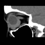
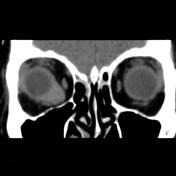
Non-contrast CT through the orbits demonstrates a homogeneous soft tissue density mass abutting the inferior and medial surface of the globe, conforming to the globe's outline. The globe itself is normal and the mass does not appear to arise from the extraocular muscles or optic nerve.
The contralateral orbit appears normal
Case Discussion
Findings are consistent with primary orbital lymphoma which was subsequently histologically proven.


 Unable to process the form. Check for errors and try again.