
Presentation
Painful left proptosis.
Patient Data
Age: 40 years
From the case:
Orbital pseudotumor


Download
Info
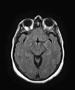
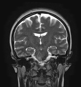
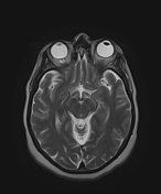
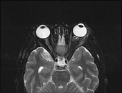
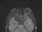
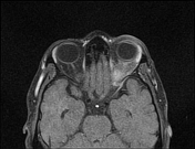
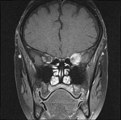
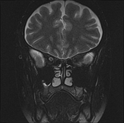
The MRI sequences demonstrate left proptosis grade I with infiltration of the retrobulbar intraconal fat of intermediate signal intensity on T1WI, low signal intensity, T2WI, and FLAIR, relatively high signal intensity on T2 fat sat sequences. The postcontrast sequences show moderate enhancement as well as an enhancement of the ipsilateral optic nerve sheath. The lateral/superior rectus muscles, as well as the lacrimal gland, are enlarged with homogeneous enhancement.
Case Discussion
MRI features suggestive of orbital pseudotumor. The main differential diagnosis includes:
- orbital lymphoma: usually presents as a progressive orbitopathy, more often bilateral with lower values on ADC and does not respond to corticosteroid
- orbital cellulitis: usually associated with a subperiosteal abscess from adjacent sinusitis or presents a previous history of trauma/dental procedure
- thyroid-associated orbitopathy (TAO): usually not painful, and spares the tendinous insertions
- granulomatosis with polyangiitis: bilateral involvement of the orbits, and PNS with associated osseous destruction


 Unable to process the form. Check for errors and try again.