
Presentation
Incidental finding.
Patient Data
Age: 45 years
Gender: Female
From the case:
Ossification of the falx with fatty marrow


Download
Info
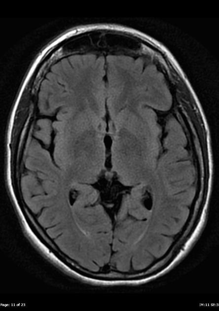
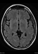
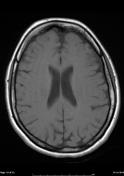
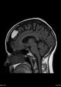
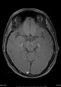
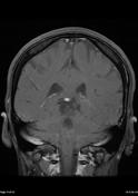
The left parafalcine/falcine mass measures 19 x 31 x 12 mm, and has internal signal consistent with fat on all sequences. It is surrounded by low signal on all sequences, almost similar presenting calcification/ossification. A similar second but much smaller region projecting on the right side of the falx, more posteriorly and superiorly, measuring only 6 x 12 x 12 mm is noted. Neither region demonstrate any abnormal enhancement, and the remainder of the brain is normal in appearance.
Appearances are characteristic, albeit pronounced, of ossification of the falx with central fatty marrow.
Case Discussion
Ossification of the falx is common and can contain marrow. When large it can be quite striking.


 Unable to process the form. Check for errors and try again.