
Presentation
Incidental
Patient Data
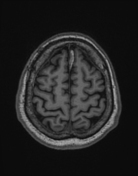

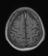
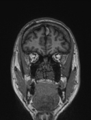
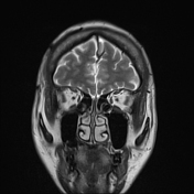
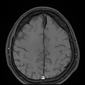


MRI shows an abnormal signal area along the anterior portion of the falx cerebri. It has a well-defined, irregular border and appears hyperintense on both T1 and T2 images at its center, with a surrounding rim of hypointensity in both T1 and T2. Also, it appears dark on SWI images, and there is no evidence of restricted diffusion.
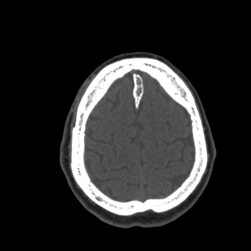
CT scan shows the same areas which hypodense in the center surrounded by a rim bone-like density
Case Discussion
The high signal observed on T1 and T2 is probably linked to the presence of marrow and/or fat components that exhibit hypodensity on CT scans. And the low signal rim detected on T1WI and T2WI, which corresponds to hyperdense areas on the CT scan, is likely cortical bone.
Therefore, an ossified falx with bone marrow represents a high signal intensity center and a periphery with low signal intensity on T1 and T2 images.
In some cases, patients with a history of head trauma may be mistaken for having a hematoma. However, based on their morphology, T1FS and CT scan results, we can clearly distinguish between the two.


 Unable to process the form. Check for errors and try again.