
Presentation
Work up for knee pain.
Patient Data
Age: 35 years
Gender: Female
From the case:
Ossifying fibroma

Download
Info
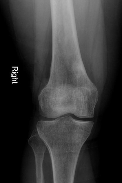
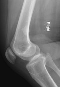
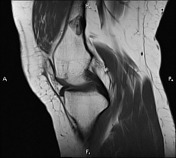
A cortical based intramedullary lesion at the distal metaphysis of the right femur. It shows small lucent core with sclerotic margin inferring ossifying fibroma. No cortical destruction is noted.
From the case:
Ossifying fibroma

Download
Info

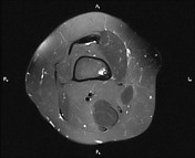
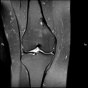
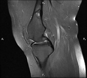
Cortical based ossifying fibroma is seen in medial aspect of distal femoral metaphysis.
Grade I intra meniscal injury is seen in posterior horn-body junction of medial meniscus.
Mild joint effusion is detected.
Case Discussion
Ossifying fibromas are benign bone lesions that should be differentiated from non-ossifying fibromas and fibrous dysplasia.


 Unable to process the form. Check for errors and try again.