
Presentation
Bilateral inguinal pain.
Patient Data
Age: 30 years
Gender: Male


Download
Info
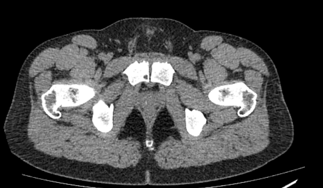
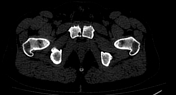
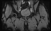
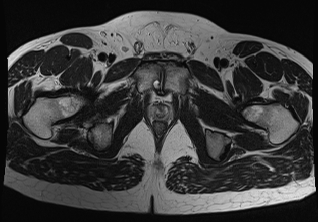
Subchondral erosive changes along symphysis pubis articular surfaces with irregular articular surfaces, subchondral sclerosis and cystic changes.


Download
Info
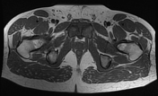
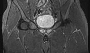
The symphysis pubis showed bilateral para-symphyseal edematous marrow changes. This is associated with feathery T2 hyperintensity at the myotendinous junction of the origin of the adductor brevis muscles bilaterally, more on the right. Intact tendons of the adductor longus muscles on both sides.
Also noted:
- bilateral Os acetabuli
- tiny subcortical cystic changes along the anterior aspect of the right femoral head-neck junction (synovial herniation pits)
Case Discussion
Osteitis pubis is inflammation of symphysis pubis and adjacent soft tissues, a cause of pelvic pain. It appears as erosive bone changes in CT and parasymphyseal bone marrow edema on MRI.


 Unable to process the form. Check for errors and try again.