
Presentation
13 year old with a large bony swelling on the mid-shin.
Patient Data


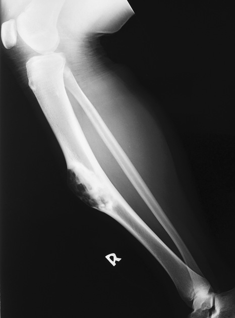
Expansile cortical based lucent lesion with a narrow zone of transition in the mid tibial diaphysis.
The anterior cortext is thinned, but not breeched. Internal 'soap bubble'appearance to the lesion which extends into the medulla and has a sclerotic rim.


T1 intermediate, T2 high signal enhancing mass in the anterior aspect of the tibial diaphysis with cortical thinning and minor breech, that extends into the medullary cavity.

Open surgical biopsy undertaken and histology report provided.
Case Discussion
Osteofibrous dysplasia is a benign bone lesion typically identified in the lower extremities. The tibia is the most frequent site accounting for 90% of all case a predilection for the anterior tibial cortex, as in this case.
The chief differentials was adamantinoma and fibrous dysplasia.
Surgical biopsy was performed to give this histologically proven diagnosis.


 Unable to process the form. Check for errors and try again.