
Presentation
Left thigh pain
Patient Data
Age: Adult
Note: This case has been tagged as "legacy" as it no longer meets image preparation and/or other case publication guidelines.
From the case:
Osteoid osteoma
Download
Info
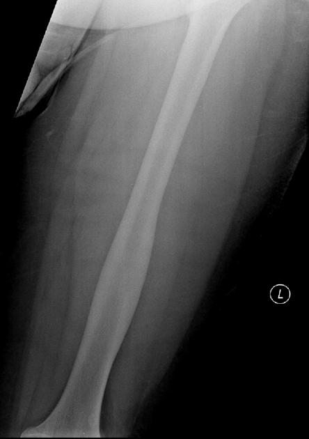
Cortical thickening and sclerosis is noted within the mid to distal femur. No periosteal reaction or other aggressive features.
From the case:
Osteoid osteoma

Download
Info
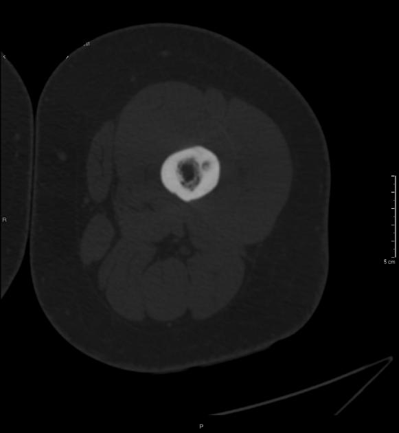
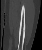
Small central lucency representing the nidus is noted within the thickened, sclerotic cortex.
Case Discussion
This lesion is an osteoid osteoma. CT is more sensitive than plain film at detecting a small nidus.


 Unable to process the form. Check for errors and try again.