
Presentation
Pain in the left leg with focal swelling in the medial aspect of proximal left leg.
Patient Data


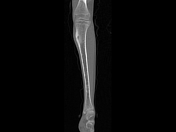
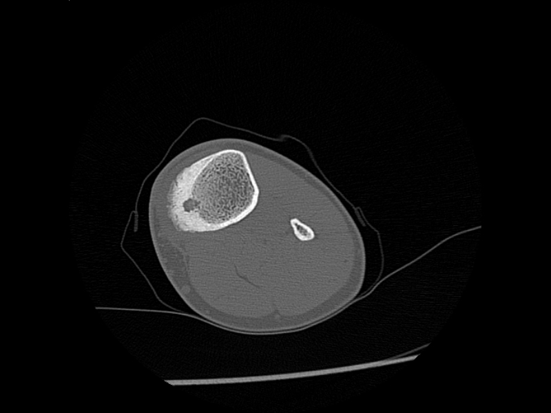
Relatively well circumscribed lucent lesion measuring approximately 28 mm long and 8 mm wide is noted involving the medial cortex of the left proximal metadiaphysis. Few tiny sclerotic dots are seen within the lesion with marked cortical thickening (13 mm) and surrounding sclerosis. No obvious soft tissue component noted. There is evidence of thin linear lucency extending from periosteal surface of the cortex down to the nidus (Vascular Groove Sign). No evidence of fracture or endosteal scalloping.
Case Discussion
The vascular groove sign is a moderately sensitive but highly specific CT feature for distinguishing osteoid osteomas from other radiolucent bone tumors. These grooves, likely corresponding to enlarged arterioles delivering blood to the hypervascular tumor nidus, align with histologic findings.


 Unable to process the form. Check for errors and try again.