
Presentation
Knee pain in immunocompromised patient on high-dose steroid therapy
Patient Data


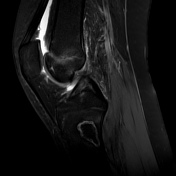
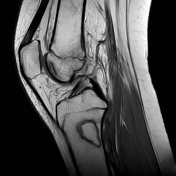
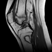
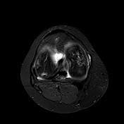
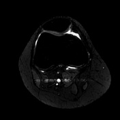

MRI shows the presence of multiple geographic regions of abnormal bone marrow signal with double line sign and rim sign, more evident in both femoral condyles, medial tibial epiphysis and proximal tibial diaphysis.
The articular surface and subchondral bone plate are subtle on the lateral aspect.
Presence of bone marrow edema surrounding the area of avascular necrosing suggests a possible impending joint collapse. Edema in the necrotic segment is seen.
These findings are in keeping with features characteristic of osteonecrosis.
Case Discussion
Osteonecrosis is a consequence of a reduction or complete loss of blood supply to the bone.
The condition has several causes and is idiopathic, the most common being trauma and heavy use of corticosteroids (as in this case) 1.
The Society of Skeletal Radiology (SSR) Subchondral Bone Nomenclature Committee prefers the term osteonecrosis to that of or avascular necrosis because all necrosis is by definition avascular 2.
Of the proposed exceptions, it prefers to speak of avascular necrosis of the knee (AVN) when the lesions are predominantly located in the epiphyses of the knee 3.


 Unable to process the form. Check for errors and try again.