
From the case:
Osteosarcoma - conventional (histology)
Download
Info
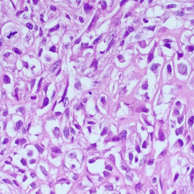
Conventional osteosarcoma histology. Image courtesy of Bulent Celasun, MD. See case description page for more details.
Case Discussion
An H&E image from a case of conventional osteosarcoma involving proximal tibia. Other areas had cartilaginous and giant cell morphology. In addition to mitosis (upper left) and cellular atypia (everywhere), which are usual; one should see the atypical osteoid (upper right) to make the diagnosis. The patient was a 14-year-old boy.
Author: Bulent Celasun, MD.
Original file: here
Modifications: square crop
License: CC BY-NC 2.0
If you believe your copyright has been infringed, please write to license@radiopaedia.org giving details of why you believe this is so.


 Unable to process the form. Check for errors and try again.