
Presentation
Further assessment of incidental finding on pelvic ultrasound, performed for vague lower abdominal/pelvic symptoms.
Patient Data
Age: Adult
Gender: Female


Download
Info
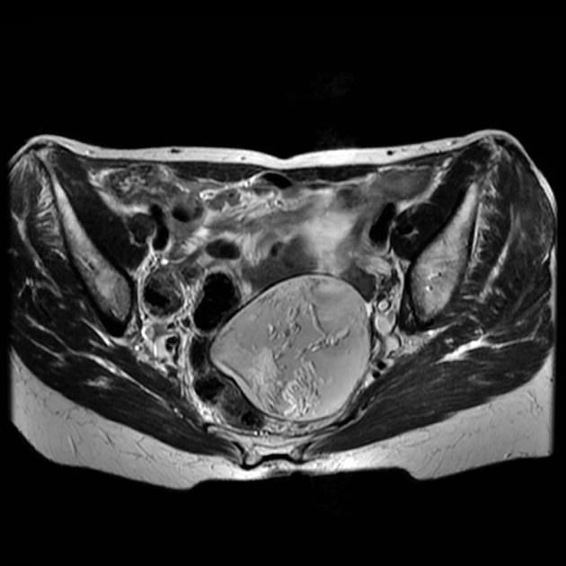
Fairly well defined heterogeneous but predominant T2 hyperintense lesion is seen in the pelvis posterosuperior to the uterus. It is predominantly of T1 hyperintense with hypointense areas within. On T2 fat-saturated images, the T1 and T2 hyperintense areas show complete signal drop. A posterior intracystic nodule is noted with mixed signal pattern suggestive of a Rokitansky nodule.
Case Discussion
This case illustrates the typical MRI features of a large ovarian dermoid with a Rokitansky nodule. This was excised and confirmed histologically.
Personal ref: V_OG 23


 Unable to process the form. Check for errors and try again.