
Presentation
Recurrent groin pain
Patient Data
Age: 40 years
Gender: Female
From the case:
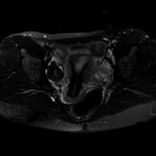
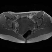
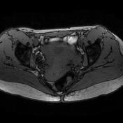
Ovarian dermoid cyst (MRI)


Download
Info
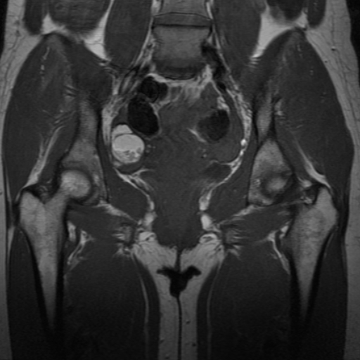
A right intra-ovarian lesion is noted with heterogeneous high signal intensity on T1WI with near-complete loss of signal on fat-suppressed T2WI. In-phase and out-of-phase sequences show no loss of signal excluding intracellular fat nature.
Case Discussion
MRI features are in favor of a fat-rich right intra-ovarian lesion with a macroscopic fat signal, in keeping with mature cystic teratoma of the ovary.


 Unable to process the form. Check for errors and try again.