
Presentation
Referred for further investigation with MRI for an abnormality detected on ultrasound
Patient Data
Age: 40 years
Gender: Female


Download
Info
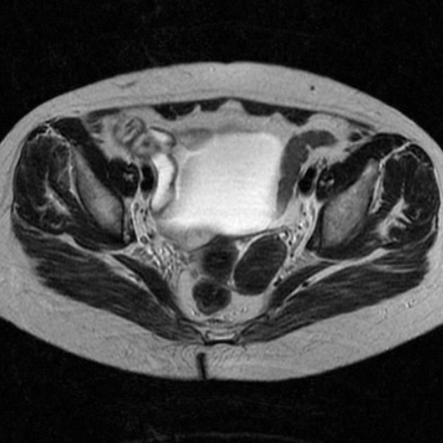
There was a hypoechoic solid mass of the left adnexa on pelvic ultrasound (not shown). This MRI was performed before laparoscopic resection.
Case Discussion
Histopathologic examination revealed a fibroma. In about 1%, these benign tumors may lead to the Meigs syndrome.


 Unable to process the form. Check for errors and try again.